You know the saying right? ‘An ounce of prevention is worth a pound of cure’. Well, nowhere is that more true than when considering dental health for our pets! This month we will be discussing the importance of dental radiography and preventive dentistry in our canine and feline patients. As with many health concerns, prevention of dental disease is far easier, less expensive and less stressful than trying to intervene after disease has taken hold. Just as most of us visit our dentist every 6-12 months, so too should our pets!
So what does preventive dentistry involve for our dogs and cats? The answer to this question will vary somewhat based on age and breed but below are general guidelines that will benefit most pets:
- 6-10 weeks of age – Visual oral and dental exam
- Most of our puppies / kittens need to be seen at this age for a general health check-up and vaccinations. This provides us with an excellent opportunity to evaluate their facial and dental conformation and start screening for problems such as malocclusions (improper alignment of teeth), delayed eruption of teeth and cleft palates. If any of these abnormalities are noted early intervention may prevent more serious problems once the adult teeth begin to erupt at about 4 months of age.
- 5-6 months of age – Visual oral and dental exam +/- anesthetized exam with full mouth intra-oral dental radiographs
- At this time all the deciduous (baby) teeth should have been lost and adult teeth are erupting. This is perhaps the most critical time for our pets with regards to their dental development. It is important that they be evaluated for proper alignment, number and appearance of their teeth. Any abnormalities such as malocclusion, retained baby teeth, missing teeth, extra teeth, crowding or abnormal morphology (appearance) warrant prompt investigation and intervention as needed. Any pet with missing teeth, crowding, malocclusion or abnormal tooth morphology needs full mouth intra-oral radiographs. The sooner these abnormalities are properly investigated and corrected the better. Once the adult teeth have finished erupting, permanent changes may have already occurred and it is much more difficult to correct these issues. For pets undergoing spay/neuter at this age it is an ideal time to get that first set of dental x-rays!
- 1-2 years of age – Comprehensive Oral Health Assessment & Treatment
- Research shows that at least 50% of our pets have established periodontal disease by 3 years of age. Since our goal is to prevent disease it is important that we start with dental care before this point. During a COHAT your pet’s veterinarian will do a detailed visual, tactile and radiographic (x-ray) assessment of your pet’s dental health. Any abnormalities found will be addressed and a customized plan for maintaining your pet’s oral health will be developed.
- 3+ years of age – Annual to biannual COHAT’s
- The frequency with which your pet will require professional dental work will vary based on breed, compliance with home-care, age and overall dental health. The more work you can do at home the less frequently your pet will need a full COHAT under anesthesia.
A Case In Point – The Dentigerous Cyst
So at this point you may be thinking to yourself that this all sounds like much ado about nothing right? Wrong. Let’s look at a recent case treated by Dr. Whitmer at Temperance Animal Hospital that illustrates just how important preventive dentistry is.
The patient is an almost 6 year old dog who was presented for treatment of an infected upper molar tooth on the other side of her mouth. When her full-mouth intra-oral dental radiographs (x-rays) were taken we discovered that an un-erupted first premolar in the bottom jaw had caused a large and very destructive cyst. These cysts can form around un-erupted teeth and as they continuously expand they destroy everything in their path including bone!
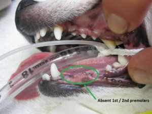
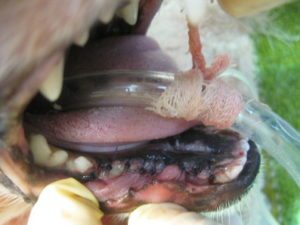
Here is a picture of her mouth before cleaning and surgery – doesn’t look too bad right? Note where there are two missing teeth on the bottom jaw. On gross examination these teeth were missing on both sides (this is the side without the cyst but they both looked the same).
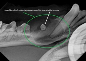
Here is a picture of her radiograph which shows the large cyst (black area) around the un-erupted first premolar.
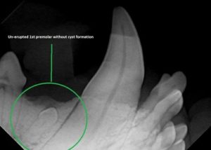
Here is a picture of a radiograph showing an un-erupted first pre-molar before a cyst has formed – notice how much bone was destroyed by the cyst in our patient’s case!
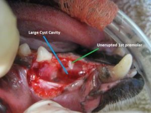
After opening up the area of the cyst we can appreciate how destructive these un-erupted teeth can become!

Unfortunately, this cyst had become so large that the surrounding canine and third premolar tooth also had to be removed. Here is a picture after removal of all the affected teeth and removal of the cyst lining.
And here is our patient’s mouth after closing the surgical wound.
Fortunately no serious complications are expected (such as jaw fracture which can result if these are left un-treated); it would have been far easier, less expensive and less traumatic to have removed this tooth at 1 year of age beforethe cyst caused so much destruction!
As part of your pet’s Veterinary Healthcare Team, we make it our mission to prevent disease whenever possible – but we can’t do this alone! You will find that dedication on your part to early and consistent attention to your pet’s oral health will pay off for years to come!